The Bruker Dimension Icon XR Atomic Force Microscope (AFM) is located in 118 AMIC at Penn State Behrend.
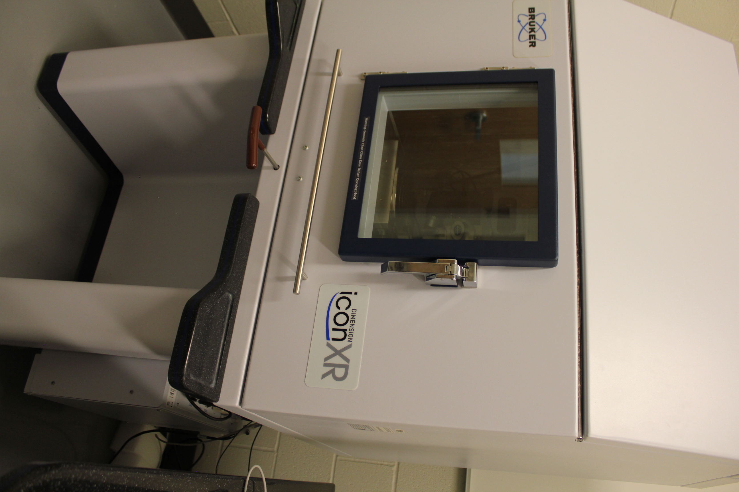
The Bruker Dimension Icon XR Atomic Force Microscope (AFM), located in 118 AMIC.
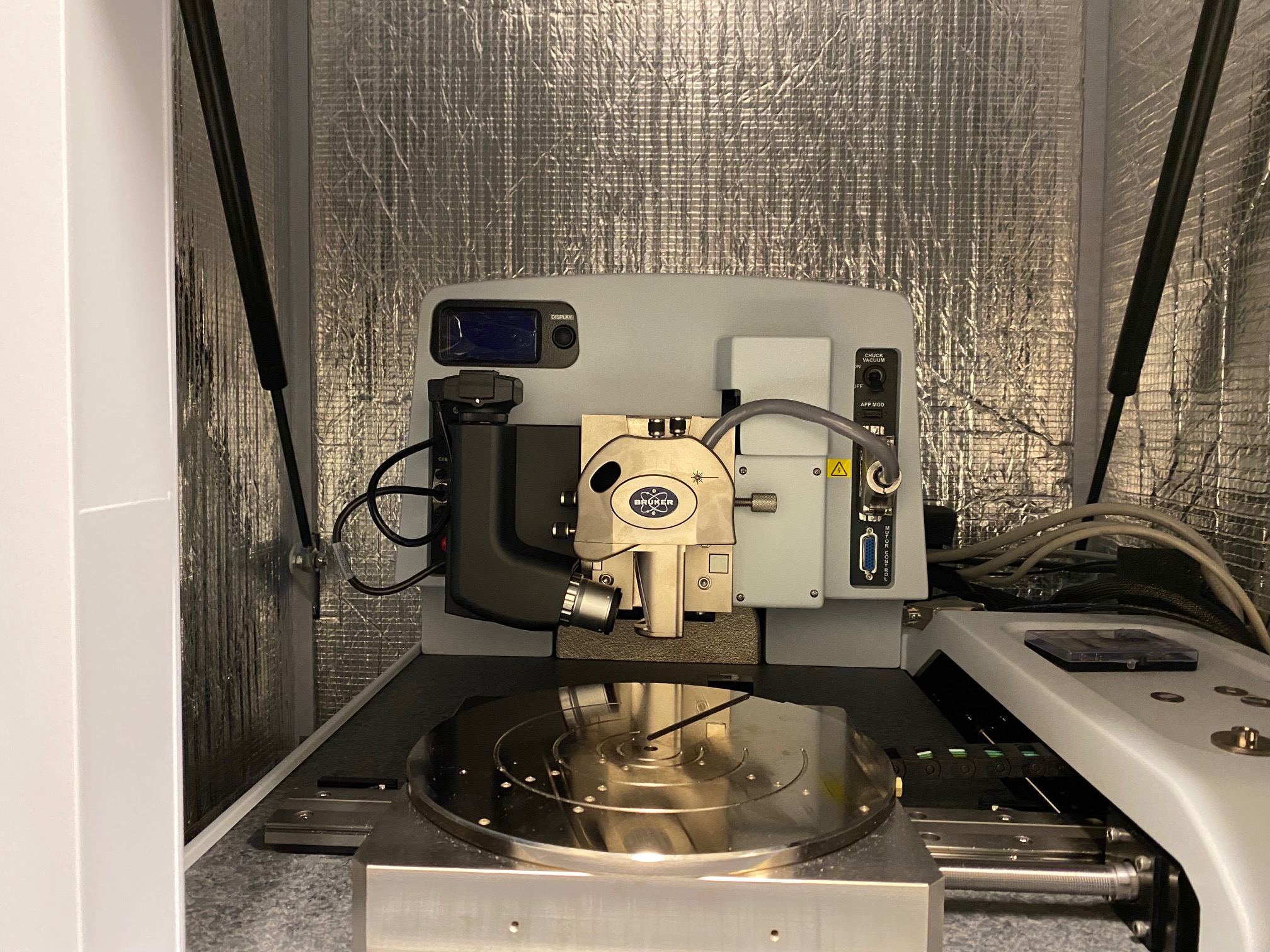
A look inside the Bruker Dimension Icon XR Atomic Force Microscope (AFM), located in 118 AMIC.
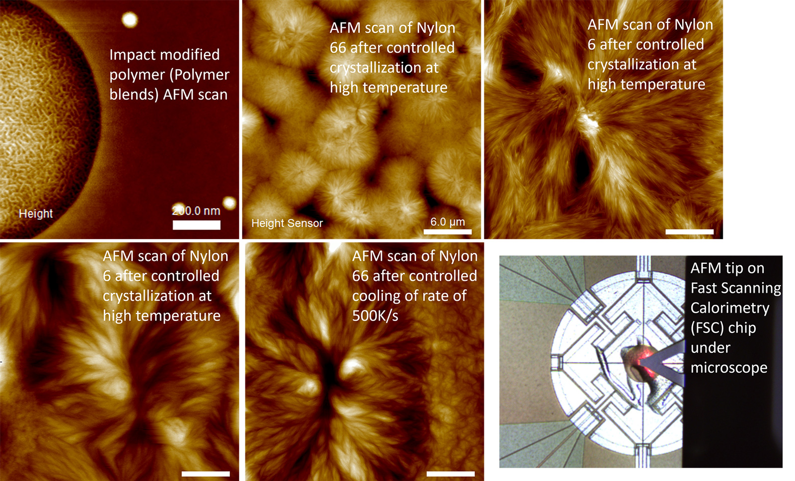
These example images show advanced research using the Bruker Dimension Icon XR Atomic Force Microscope (AFM), located in 118 AMIC.
Dr. Xiaoshi Zhang conducted AFM on a fast scanning calorimetry (FSC) chip. All samples were previously prepared on the chip with controlled crystallization pathways. The selected crystallization pathways allow us to mimic real-life processing and the AFM scan enables us to know nanoscale morphology.
Text in images (L-R):
- Impact modified polymer (Polymer blends) AFM scan
- AFM scan of Nylon 66 after controlled crystallization at high temperature
-
AFM scan of Nylon 6 after controlled crystallization at high temperature
-
AFM scan of Nylon 6 after controlled crystallization at high temperature
-
AFM scan of Nylon 66 after controlled cooling of rate of 500K/s
-
AFM tip on Fast Scanning Calorimetry (FSC) chip under microscope